Home
/ Diagram Of Animal Cell Membrane - Diagram Showing Anatomy Of Animal Cell Stock Vector Illustration Of Nature Drawing 74438191 / In the labeled animal cell diagram, it is nearly circular in shape and lacks outer cell wall;
Diagram Of Animal Cell Membrane - Diagram Showing Anatomy Of Animal Cell Stock Vector Illustration Of Nature Drawing 74438191 / In the labeled animal cell diagram, it is nearly circular in shape and lacks outer cell wall;
Diagram Of Animal Cell Membrane - Diagram Showing Anatomy Of Animal Cell Stock Vector Illustration Of Nature Drawing 74438191 / In the labeled animal cell diagram, it is nearly circular in shape and lacks outer cell wall;. Cells are covered by a cell membrane and come in many different shapes. Structure of membrane in prokaryotes cell: These are organelles pertinent to plant cells. Drawing of the fluid mosaic model. That cells can be of different shapes and sizes.
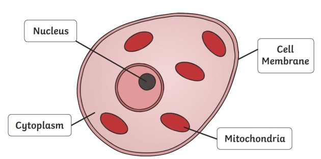
The ones mentioned on this page include centrosomes, goli apparatus, lysosomes, mitochondria, the nucleus and its parts such as the nuclear membrane and nuclear pores, also other. An animal cell diagram is a great way to learn and understand the many functions of an animal cell. It is the outermost part of the cell in animals. The diagram, like the one above, will include labels of the major parts of an animal cell including the cell membrane, nucleus, ribosomes, mitochondria, vesicles, and cytosol. The contents of a cell are called the protoplasm.
What Is An Animal Cell Answered Twinkl Teaching Wiki from images.twinkl.co.uk The diagram is very clear, and labeled; However, in plants, bacteria, and fungi, it is surrounded by a thick cell wall. A vast system of interconnected, membranous, infolded and convoluted sacks that are located in the cell's cytoplasm (the er is continuous with the outer nuclear membrane). Animal cells, plant cells, prokaryotic cells, and fungal cells have plasma membranes. Here's a diagram of a plant cell: The chapter is number 5 titled membrane structure and function. Note that they are composed of phospholipid molecules and protein. Internal organelles are also encased by membranes.
Animal cells, plant cells, prokaryotic cells, and fungal cells have plasma membranes.
Structure of membrane in prokaryotes cell: They are different from plant cells in that they do contain cell walls and. Draw a table of differences between the two cell types in the space provided. Removing cellular waste products from the cell. The diagram, like the one above, will include labels of the major parts of an animal cell including the cell membrane, nucleus, ribosomes, mitochondria, vesicles, and cytosol. Animal cell membrane diagram page for you to see. After completing this section, you should know: This organelle is also referred to as plasma membrane. Diagram of plasma membrane (cell membrane), created with biorender.com. The cell membrane (or plasma membrane) is the thin outer layer of the cell that differentiates the cell from its environment. Cell membranes, also called the plasma membrane, is a physical barrier between a cell and the surrounding environment. Smooth endoplasmic reticulum, mitochondria, golgi bodies, lysosomes. Cytoplasm, ribosomes, rough endoplasmic reticulum;
Solved label the parts of a cell membrane with the term. Internal organelles are also encased by membranes. Animal cells, plant cells, prokaryotic cells, and fungal cells have plasma membranes. Cholesterol is a component of animal cell membranes. Unlike the eukaryotic cells of plants and fungi, animal cells do not have a cell wall.
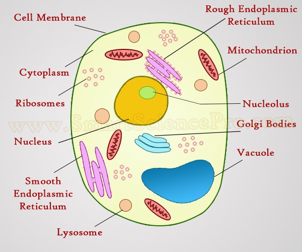
Structure Of Animal Cell And Plant Cell Under Microscope Diagrams from www.smartsciencepro.com However, in plants, bacteria, and fungi, it is surrounded by a thick cell wall. Unlike the eukaryotic cells of plants and fungi, animal cells do not have a cell wall. All animal cells contain organelles. In plant cells, peroxisomes play a variety of roles including converting fatty acids. Essentially, a cell membrane is the outermost barrier that separates the internal contents of a cell in the cytoplasm from the external environment (e.g. After completing this section, you should know: Removing cellular waste products from the cell. Cholesterol in mammalian membranes reduces membrane fluidity and permeability to some solutes.
Draw a table of differences between the two cell types in the space provided.
Animal cell functions are solely dependent on the organelles and structures associated with the cell. Below is a diagram of a part of the plasma membrane. A cell (plasma) membrane encloses the cytoplasmic contents, such as nucleus, peroxisome, cytoskeleton, lysosome, ribosomes, mitochondria. A simple animal cell definition. The cell membrane (or plasma membrane) is the thin outer layer of the cell that differentiates the cell from its environment. Structure of membrane in prokaryotes cell: Cell membranes, also called the plasma membrane, is a physical barrier between a cell and the surrounding environment. Smooth endoplasmic reticulum, mitochondria, golgi bodies, lysosomes. Essentially, a cell membrane is the outermost barrier that separates the internal contents of a cell in the cytoplasm from the external environment (e.g. This organelle is also referred to as plasma membrane. It is the outermost part of the cell in animals. 4) cell membrane or plasma membrane. Cholesterol is a component of animal cell membranes.
Start studying label cell membrane. In animals, the cell membrane establishes this separation alone, whereas in yeast, bacteria and plants. The chapter is number 5 titled membrane structure and function. Solved label the parts of a cell membrane with the term. The animal cell is made up of several structural organelles enclosed in the plasma membrane, that enable it to function properly, eliciting mechanisms that benefit the host (animal).
Free Vector Human Cell Membrane Structure Illustration from img.freepik.com Essentially, a cell membrane is the outermost barrier that separates the internal contents of a cell in the cytoplasm from the external environment (e.g. Smooth endoplasmic reticulum, mitochondria, golgi bodies, lysosomes. Animal cell functions are solely dependent on the organelles and structures associated with the cell. Start studying label cell membrane. Most prokaryotic cells have only the plasma membrane. Internal organelles are also encased by membranes. Animal cells, plant cells, prokaryotic cells, and fungal cells have plasma membranes. The animal cell is made up of several structural organelles enclosed in the plasma membrane, that enable it to function properly, eliciting mechanisms that benefit the host (animal).
Animal cells, plant cells, prokaryotic cells, and fungal cells have plasma membranes.
Below is a diagram of a part of the plasma membrane. Draw a table of differences between the two cell types in the space provided. Schematic diagram of a cell membrane. A cell (plasma) membrane encloses the cytoplasmic contents, such as nucleus, peroxisome, cytoskeleton, lysosome, ribosomes, mitochondria. Cholesterol is another lipid component of animal cell membranes. It is the outermost part of the cell in animals. The cell membrane (or plasma membrane) is the thin outer layer of the cell that differentiates the cell from its environment. They are different from plant cells in that they do contain cell walls and. These are organelles pertinent to plant cells. The diagram, like the one above, will include labels of the major parts of an animal cell including the cell membrane, nucleus, ribosomes, mitochondria, vesicles, and cytosol. A vast system of interconnected, membranous, infolded and convoluted sacks that are located in the cell's cytoplasm (the er is continuous with the outer nuclear membrane). The cells of animals are. Study the two diagrams of plant and animal cells below.
Share :
Post a Comment
for "Diagram Of Animal Cell Membrane - Diagram Showing Anatomy Of Animal Cell Stock Vector Illustration Of Nature Drawing 74438191 / In the labeled animal cell diagram, it is nearly circular in shape and lacks outer cell wall;"


Post a Comment for "Diagram Of Animal Cell Membrane - Diagram Showing Anatomy Of Animal Cell Stock Vector Illustration Of Nature Drawing 74438191 / In the labeled animal cell diagram, it is nearly circular in shape and lacks outer cell wall;"